ORACLE: June 7, 2019
June 7, 2019
Today’s paper is, “Simultaneous mesoscopic and two-photon imaging of neuronal activity in cortical circuits”, by Barson D, Hamodi AS, Shen X, Lur G, Constable RT, Cardin JA, Crair MC & Higley MJ. We read this in our lab meeting, and James Roach took the lead on writing it up.
This article brings together two powerful experimental approaches for calcium imaging of cortical activity: 1) using viral injections to the transverse sinus to achieve high GCaMP expression throughout the cortex and thalamus and 2) using a right angle prism and two orthogonal imaging paths to simultaneously capture mesoscale activity from the dorsal cortex and 2-photon single cell activity.
Big Question: How diverse is the cortex-wide functional connectivity of neurons within a local region of the cortex and do these patterns depend on cell type?
Approach: The authors performed mesoscale  calcium imaging paired with 2-photon imaging from the primary somatosensory cortex (S1) in awake and behaving mice. Leveraging the novel technical approaches that they introduce here (and [1]), the authors quantify the relationship between the activity of individual cells and populations across the cortex. Using a method similar to spike-triggered-averaging (the cell centered network; CCN), they show that the activity-defined correlations patterns of S1 pyramidal neurons are super diverse (right*). Interestingly, neurons with similar cortex-wide functional connectivity are not necessarily spatially organized in S1.
To examine whether linkages to cortical networks differ based on cell type and behavioral role, animals expressing td-tomato in VIP+ neurons were injected pan-neuronally with GCaMP. VIP+ neurons were far more homogeneous in responsiveness to whisking and running behaviors than presumptive pyramidal cells. For both neuron types, membership in cortical networks was predicted by whether the cell was correlated with whisking or running behaviors. These combined effects lead to VIP+ neurons being far less diverse than non-VIP+ neurons in functional cortical connectivity.
Main take-home: Within a small patch of cortex, the diversity of functional relationship neurons form across the cortex is surprisingly high and that the local spatial arrangement of neurons is not in a “corticotopic map”: two neighboring cells can differ greatly in functional connectivity. The behavioral tuning of a neuron determines, or is the result of, its membership within a functional network. VIP+interneurons have a reduced diversity of functional connectivity, but this may be a result of VIP+ neurons being more similarly tuned to running or whisking.
Methodologically, this paper makes a significant advance in multiscale recording of neural activity. Pairing 2-photon and mesoscale calcium imaging provides a meaningful advantage in that neuronal cell types can be genetically targeted, cells can be recorded from stably for multiple days, and meso- and microscale activity can be recorded from the same brain region. We were quite impressed by how good the signals from both modalities were (although we would love to see more data comparing the two, especially pixel-by-pixel correlations). Plus, viral injections into the transvers sinus provide for high expression levels without the drawback of other possible sites.
Skeptics Corner: First, we were a bit concerned that the functional connectivity analysis loses a lot of detail after significance thresholding of the cell centered network data. Preserving some of the complexity in the raw CCN values might support a better alignment to AIBS-defined brain regions. For example cell 2 in figure 3d (below*) shows peaks in CCN in sensory and motor areas separated by regions with lower correlations, but thresholding for significance leads to these areas being treated equally.
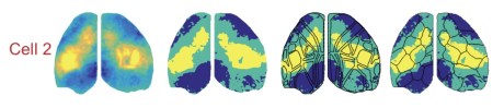
Second, many of the results depend on the functional parcellation of the cortex based upon mesoscale data and we’d love to know a lot more about the outcome of alternative parcellation strategies. Sixteen parcels per hemisphere was chosen to match to number of regions in the Allen CCF, but how does this parameter affect the analysis? An alternative method, Louvain parcellation (Vanni et al., 2017), does not require the user to specify number of clusters in advance, so we were curious about what that would look like. Also, presenting the functional parcels color-coded for modality implies a bilateral symmetry which is not reliably supported by the borders of the regions (i.e. the hemispheres in figure 3e would look quite different without the color coding). Quantifying the bilateral symmetry of the parcel boundaries would be useful both for imposing a cost for deviating from symmetry, but also for identifying states or conditions which lead to lateralized cortical activity.
Third, when comparing VIP neurons to putative pyramidal, the distinction of GCaMP+/TdT+ for VIP and GCaMP+/TdT- for pyramidal excludes the possibility that non-VIP interneurons could be expressing GCaMP. Might this contribute to the observed increase in functional diversity for pyramidal neurons reported?
Finally, reporting the cortex-wide functional relationships of the neurons that are not modulated by running or whisking would be an interesting result to add. Are these cells a diverse subset of the S1 population or are they a single functional block?
Outlook: This paper builds on results that highlight the functional diversity of cortical neurons within local circuits. The indication that cell-to-cortex functional relationships can be modulated by behavior highlight that shaping how individual neurons interact with brain wide networks is a central feature of brain states. Multiscale neurophysiology will be a crucial tool in establishing these relationships. An intriguing morsel of information in the article is this: while cell-spiking was uncorrelated with mesoscale activity at a given location, summed spiking and neuropil signal were. This will be important when interpreting a brain region’s mesoscale signal as representing the inputs to, outputs from, or a combination of the two. Mesoscale recordings with soma-targeted calcium reporters (once available) will be useful in disentangling the components of the signal.
*Note we present figure panels from the manuscript with slight modification (cropping) in accordance with the CC BY-NC-ND 4.0 license.
[1] Hamodi A, Martinez Sabino A, Fitzgerald ND, Crair MC (2019) Transverse sinus injections: A novel method for whole-brain vector-driven gene delivery. BioRxiv.
[2] Vanni, M.P., Chan, A.W., Balbi, M., Silasi, G., and Murphy, T.H. (2017). Mesoscale Mapping of Mouse Cortex Reveals Frequency-Dependent Cycling between Distinct Macroscale Functional Modules. J Neurosci37, 7513-7533.
