H. Read identifies auditory areas by intrinsic imaging & measures responses electrophysiologically to define function
October 31, 2014
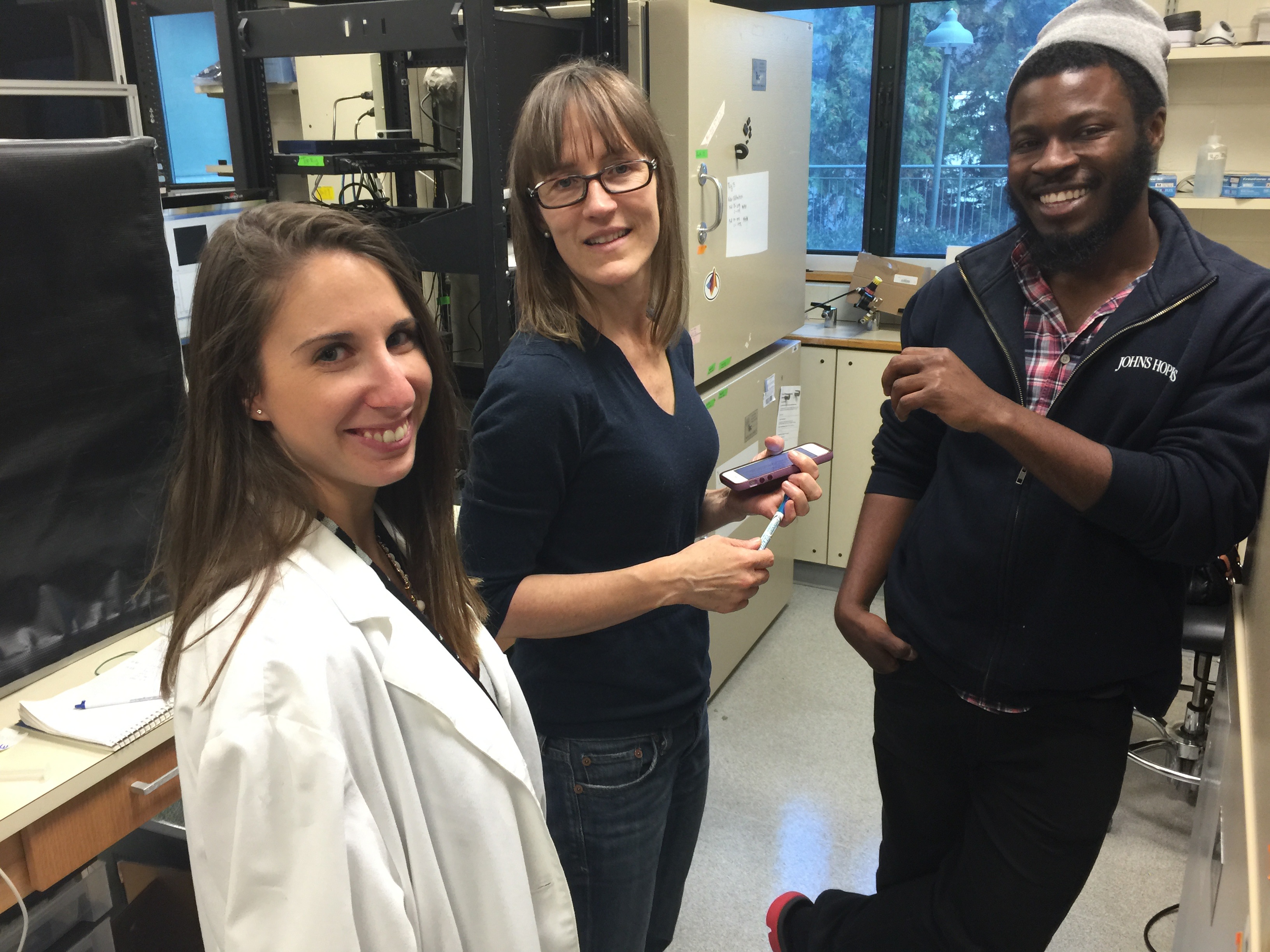 Heather Read, from the University of Connecticut, visited my lab this week. She shared her expertise on intrinsic imaging with my lab members and I, and had extensive conversations with Kachi Odeomene and Lital Chartarifsky (shown at left). Heather also gave a seminar describing her recent work measuring neural responses in 3 auditory areas. Heather and her colleagues are interested in how these areas work together to provide the needed information about auditory inputs to make sense of them. The first thing they do is to use intrinsic imaging to map out the three auditory areas (see image below). To do this, they take advantage of the fact that each area has a unique “map” of tonotopic space. But why would the brain need so many maps of the
 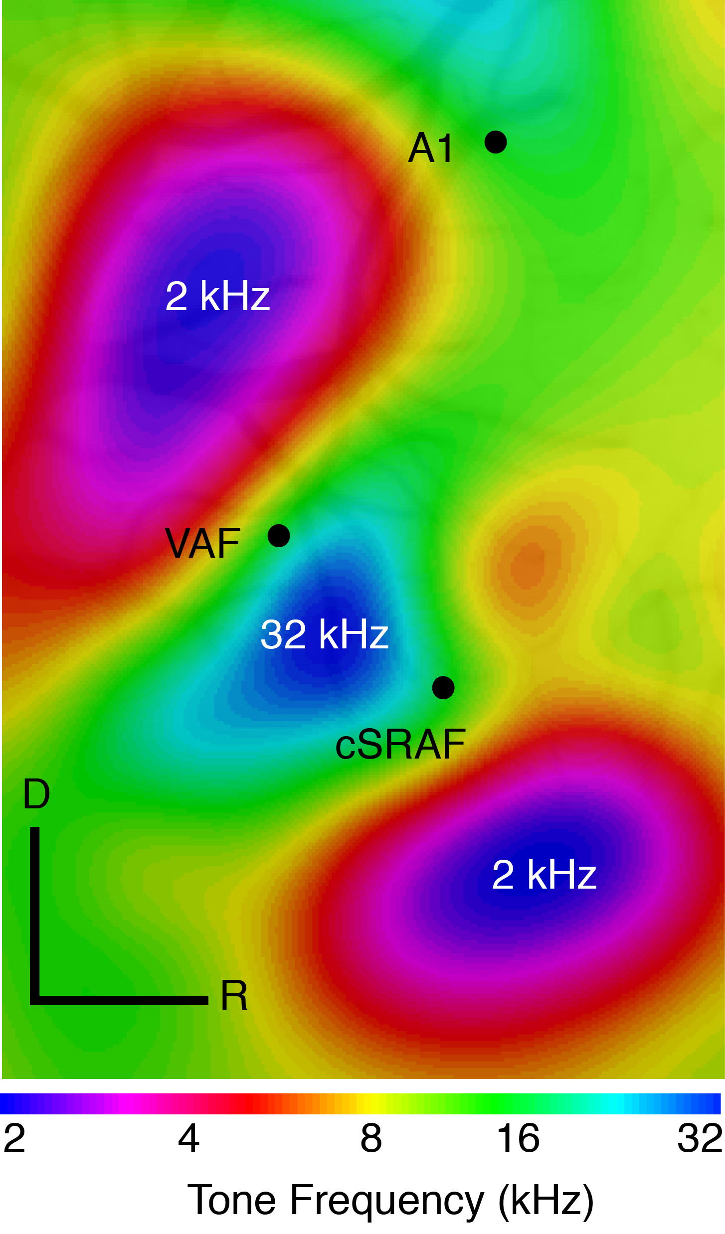 same space? One possibility is that each is specialized for a particular “shape” of sound. That is, the time and frequency modulation pattern that make a sound unique. Heather reports that three areas, primary auditory cortex, the ventral auditory field and the supra rhinal auditory field differ in the degree to which they are specialized for representing fast-modulating vs. slow-modulating sounds. This has many parallels to the visual system where visual areas differ tremendously in the degree to which they reflect fast versus slow modulating inputs.
