In this paper, Zhao and Kording aim to offer insight into the best model to describe the responses of LIP neurons during a perceptual decision-making task.
Approach:
The authors fit 3 models to LIP data from the Roitman dataset.

Model 1: “constant” or “baseline” model in which the authors attempted to model the data by a constant firing rate that fluctuates from trial-to-trial (top panel). A clarification: although this model is sometimes referred to in the paper as a “baseline” model, this doesn’t refer to activity during the time before the trial starts. Instead, it reflects that the constant that is added to the GLM; there is a single constant for the whole trial.
Model 2: “Stepping” model , in which the data jumps from a low to a high state and a time that varies trial-to-trial.
Model 3: “Ramp” model in which a linear, time dependent rise is used. Its slope can vary trial-to-trial, but all ramps start at zero. A few details: (1) This model doesn’t actually reflect a true candidate hypothesis about LIP activity: the atual hypothesis is that the activity reflects a random walk (Diffusion model), the average over many instances will approximate a ramp. However, without knowing the actual incoming evidence on the trial (not known for this dataset), modeling the random walk isn’t possible. (2) Although the models schematically depict the model the way I have drawn it here (see Fig 2B), the authors actually implemented this as an exponential ramp (I am not sure why they chose this parameterization).
Main take-home, as Zhao and Kording see it: The “constant” model fit the data best. If true, this would suggest an entirely different view of LIP activity. Specifically, not only would it suggest that neither steps nor random walks underlie the neural activity, but also that there is no time-varying change in firing rate in LIP neurons during decision-making.
Skeptics Corner:
- We had some concerns about the method for model assessment. These are nicely summarized in another blog about the paper.
- The selected model will not account for the trial-averaged data (nor the VarCE, a measure of spike-count variability [1]). To be concrete, the trial-averaged firing rate (and VarCE) progressively increases during decision-formation (a ramp) but the model is a flat line. The authors acknowledge this, but fail to explain why. In our view, the fact that the constant model was the best fit for single trial activity, but fails to explain the average, exposes a critically important truth about neural activity: modulation of neural activity due to cognitive processes (like decision-making) is strongly affected by fluctuations that arise from other neural processes, which we might refer to as “Internal Backdrop“. These other processes likely include a number of things: for instance the animal’s overall state of arousal will vary quite a bit trial-to-trial, leading the baseline firing rate to fluctuate up and down. Accounting for this baseline activity is challenging, but possible [2], and allows an investigator to separate components of the neural signal due to the internal backdrop of brain activity from decision-related activity.
- An easy way around this problem is
 o include a fourth model, perhaps termed “Ramp with trial-to-trial variation” [2]. I know it isn’t a great name. I am not famous for picking catchy names (remember the VarCE?). This would allow for the reality that decision-related signals ride on top of trial-to-trial variability in baseline firing rate and should outperform models that only account for ramps or constant changes. The authors actually did mention such a model in passing, but stated that it did worse than the model with only baseline fluctuations which at first seems odd (how could adding a parameter worsen the fit?). My hypothesis is that the failure of the model with more parameters stems from overfitting. Here’s why: they are fitting individual time bins (again, see this blog) and then cross validating on left out time bins. However, estimating a ramp in a small time bin is very challenging. The ramps are meant to unfold slowly over the whole trial (and they aren’t ramps on single trials at all). Plus, the point process variance will prevent one from getting a meaningful estimate of a ramp at all. So of course, much of the time one would estimate the wrong ramp, leading to bad predictions on the left-out time bins and hence the poor model fit.
 - The Roitman dataset (available here) was appropriate in some ways, most notably that it was the dataset analyzed in a related paper examining this issue [3]. However, this dataset was ill-suited to some of the analyses in the paper because there are low trial counts for many neurons (large trial counts are critical because there are 10 conditions: 2 motion directions and 5 coherence levels). This is already a challenge when estimating firing rate mean, and will be an enormous problem when estimating firing rate variance, as was done to compute the Fano Factor in Figure 1. The paper reports FFs as large as 8, which, while not impossible, likely result from an uncertain estimate of spike count variance.
- Finally, the Roitman dataset only includes information about average stimulus coherence for each trial; the motion energy on individual trials is missing. This prevents the possibility of actually modeling the random walk (Model 4) that is the true alternative hypothesis to the baseline and stepping models. A better test of this hypothesis could be made using stimuli for which stimulus strength is explicit such as in this paper or this paper.
Outlook
The authors emphasize the importance of simple models and highlight the importance of trial-by-trial variability when explaining data variance. We agree with this notion and believe that explanations of neural dynamics during decision-making must take the trial-by-trial internal backdrop into account. We hope that the authors will therefore consider models that combine trial-to-trial baseline variability with stimulus-evoked dynamics. A fruitful avenue for model comparison would also be to use more than only the Roitman dataset; while the authors recorded what was at the time a very large number of neurons, there are other, larger datasets available that may make it easier to arbitrate between models.
References
1. Churchland, A. K. et al. Variance as a signature of neural computations during decision making. Neuron 69, 818-831, doi:10.1016/j.neuron.2010.12.037 (2011).
2 Musall, S., Kaufman, M. T., Gluf, S. & Churchland, A. K. Movement-related activity dominates cortex during sensory-guided decision making. BiorXiv (2018).
3 Latimer, K. W., Yates, J. L., Meister, M. L., Huk, A. C. & Pillow, J. W. Single-trial spike trains in parietal cortex reveal discrete steps during decision-making. Science 349, 184-187, doi:10.1126/science.aaa4056 (2015).
Today’s commentary brought to you from (in alphabetical order): Lital Chartarifsky, Anne Churchland, Ashley Juavinett, Farzaneh Najafi, Anne Urai, & Sashank Pisupati. Feel free to comment, correct, express skepticism, etc. You can do so here, on Biorxiv or on Twitter (@anne_churchland). Let’s get a conversation going!
Paper #1: Psychophysical reverse correlation reflects both sensory and decision-making processes (Okazawa, She, Purcell & Kiani)
Big question: Psychophysical kernels are a powerful method to derive the spatiotemporal filter that transforms sensory information into a decision. However, can psychophysical kernels be interpreted as reflecting such sensory weighting profiles when measures in realistic decision-making scenarios?
Summary: First, tasks with a fixed stimulus duration cannot correctly retrieve sensory filtering timecourses, since the temporal weighting function may just as well reflect the process of bound-crossing during evidence accumulation (and the experimenter doesn’t have access to the time of the decision). Second, variable non-decision time (even in a reaction time task) results in decaying kernels. The authors then demonstrate different ways to draw informative conclusions from psychophysical kernels. First, they compare kernels in a motion direction discrimination RT task to explicit predictions derived from the DDM, and show that kernel shape can be predicted by a DDM with stationary sensory weights. The investigate a range of models that all have stationary sensory weights, and show (Figure 7) that these can generate a diversity of kernel dynamics.
Take home: Be very careful when interpreting psychophysical kernels as reflecting purely sensory weights!
Paper #2: Cortical neural activity predicts sensory acuity under optogenetic manipulation. John J. Briguglio, Mark Aizenberg, Vijay Balasubramanian, Maria N. Geffen (Note that its now in J. Neurosci).
Big question: Why does stimulation (optical, chemical, electrical) cause idiosyncratic changes in behavior, sometimes in opposing directions?
Take home: Behavioral variability occurs because the changes to neurons are also variable. In this paper, the authors showed that changes in psychophysical threshold following A1 optogenetic stimulation were variable and that this variability could be understood if one took into account the change in the neurometric threshold at the site of stimulation.To estimate neurometric threshold, the authors recorded neural activity at the same sites where they stimulated and they measured neurometric threshold using Fisher information. To estimate behavioral threshold, the authors used a pre-pulse inhibition task in which an auditory tone, if the animal’s heard it, could ward off a startle reflex in response to a loud white noise burst.
Skeptics’ corner: Changes in psychometric functions were related to changes in neurometric functions (cool!), but I was left wondering why the changes in neurometric functions were idiosyncratic. The direction of the change in neurometric threshold was idiosyncratic across sites, and even across stimulation methods. In other words, the same kind of stimulation (e.g., ChR2 stimulation in pyramidal neurons) sometimes made both neurometric threshold go up, and other times made it go down.
Paper #3: Functional selectivity and specific connectivity of inhibitory neurons in primary visual cortex Petr Znamenskiy, Mean-Hwan Kim, Dylan R. Muir, Maria Florencia Iacaruso, Sonja B. Hofer, and Thomas D. Mrsic-Flogel
Question: Do inhibitory neurons connect broadly to all their nearby excitatory neurons? Or is there specific connectivity in the connection of inhibitory to excitatory neurons?
Take home message: Inhibitory neurons connect more strongly to nearby excitatory neurons with similar responses to visual stimulation, suggesting that connections between inhibitory and excitatory neurons are organized under a similar rule to excitatory-excitatory connections. In more detail, although inhibitory neurons are less tuned to visual stimuli than excitatory neurons, their response selectivity is not merely a reflection of their surrounding neurons: inhibitory neurons selectivity out-performs that of their surrounding neurons. This is due to their selective connectivity to excitatory neurons with similar tuning properties.
Skeptics’ corner: If there is selective connectivity between excitatory and inhibitory neurons, why are inhibitory neurons still less tuned? 2) Do the same conclusions apply to other subtypes of inhibitory neurons?
Paper #4: Stable representation of sounds in the posterior striatum during flexible auditory decisions (Guo, Walker, Ponvert, Penix, Jaramillo)
Big question: What is the role of posterior striatum during auditory-driven decisions in mice?
Take home message: Posterior striatum (also known as “auditory striatum”) is causal in an auditory discrimination task.Bilateral muscimol inactivation of this area impaired performance, while unilateral optogenetic activation during sound presentation biased the animals’ choices contralaterally. The authors also showed that the activity of neurons in posterior striatum reliably encoded stimulus features, but were minimally influenced by the animals’ choices, suggesting that neurons in the posterior striatum provide sensory information downstream, while providing little information about behavioral choice.
Skeptics’ corner: The result showing impaired performance after bilateral muscimol inactivations was averaged across 4 sessions, however the authors note that on individual sessions the mouse was idiosyncratically biased to either the left or the right side. This side bias was probably caused by unbalanced muscimol injection. This is something that we should be mindful of when interpreting performance after bilateral manipulations.
Paper #5: Limitations of proposed signatures of Bayesian confidence
(William T. Adler, Wei Ji Ma)
Big question: A bayesian model of confidence was previously proposed by Hangya et. al, in which confidence reflects the subject’s estimate of posterior probability of the chosen option. Do the proposed signatures of bayesian confidence generalize?
Take home: Proposed signatures of bayesian confidence (i.e. divergence of mean confidence as a function of stimulus magnitude on correct and error trials, mean confidence of 0.75 on uninformative trials) are not necessary if the category-conditioned stimulus distributions are overlapping, especially in certain noise regimes, and yet others can be predicted by non-bayesian models. Hence favor model comparison over signatures!
Skeptics’ corner: The authors mention that an alternate model of confidence, the distance of an observation from the category boundary, can account for some of the signatures. However the question remains whether the Bayesian model makes unique predictions that distinguish it from alternatives, for instance predicted effects of changing subjects’ priors. Enumerating such unique predictions would help in directly testing the model experimentally, and ease the burden on model comparison.
ORACLE: Options to Read in the Archive: Churchland Lab’s Experience
January 31, 2018

Today’s commentary brought to you from (in alphabetical order): George Bekheet, Lital Chartarifsky, Anne Churchland, Ashley Juavinett, Simon Musall, Farzaneh Najafi & Sashank Pisupati. Feel free to comment, correct, express skepticism, etc. You can do so here, on Biorxiv or on Twitter (@anne_churchland). Let’s get a conversation going!
Paper #1: Discrete attractor dynamics underlying selective persistent activity in frontal cortex (,,
Big question: What is the deal with the persistent activity in mouse area ALM that precedes licking movements?
Take home: Using intra and extracellular recording, combined with optogenetics and network modeling (nice!), the authors conclude that attractor dynamics, and not integration, define neural activity in area ALM.
But, hmmmm: Persistent activity in advance of movements is widely observed in many critters, but its function is pretty mysterious. Other kinds of persistent activity, like memory or evidence accumulation have a clear cognitive function, but the role of motor preparatory activity is not obvious. Why does ALM need to respond to far in advance of a movement anyway?
Paper #2: Optogenetically induced low-frequency correlations impair perception (Nandy, Nassi1 & Reynolds)
Take home: Using depolarizing opsin C1V1 in a lentivirus in combination with an artificial dura, the researchers created a preparation in which they could optically excite specific locations of pyramidal cells in V4. During an orientation-change detection task, they use this system to induce low frequency (4-5 Hz) as well as high frequency (20 Hz) oscillations within the receptive field of the attended region. They demonstrate that low frequency stimulation within the attended field impairs the animal’s ability to do the task, whereas low frequency stimulation in an unattended field does not. The finding was also frequency specific — high frequency stimulation does not impair performance.
But, hmmmm: These findings provide nice closure to previous skepticism that changes in correlation structure could simply be an off-target effect, but not actually causal for attention. Still, the field seemed pretty convinced that low frequency correlations were somehow involved in attention, so this result probably will not shock many researchers. In addition, the behavioral effects are not well characterized in this paper. The two sample sessions in Figure 2, Supplement 2 show very different effects on the psychometric curve — one is shifted, whereas one has a different slope, suggesting different underlying impairments. We’d love to see the researchers more closely quantify the impairments in each animal.
Paper #3: Exclusive functional subnetworks of intracortical projection neurons in primary visual cortex (,, , &
Big question: How do long-range projection targets constrain local connectivity of cortical neurons?
Take home: distinct populations in V1 project to higher visual areas AL and PM. These distinct populations avoid making connections with each other which is unexpected given their signal correlations (response similarity). Therefore, projection target acts independently of response similarity to constrain local cortical connectivity. The absence of recurrent connections between AL and PM potentially allows for their independent modulation by top-down signals.
But, hmmmm: Should we worry that retrograde labeling may have failed to label all projections neurons? Also is identifying double labeled neurons an error-prone task?
Paper #4 Accurate Prediction of Alzheimer’s Disease Using Multi-Modal MRI and High-Throughput Brain Phenotyping (Wang, Xu, Lee, Yaakov, Kim, Yoo, Kim & Cha)
Paper #5: Causal contribution and dynamical encoding in the striatum during evidence accumulation (Yartsev, Hanks, Yoon & Brody)
Big question: Which regions of the brain are causally involved in evidence accumulation during decision making?
Take home: Anterior dorsal striatum satisfies 3 major criteria for involvement, as revealed by a detailed behavioral model – necessary (pharmacological inactivation makes accumulation noisy), represents graded evidence on single trials (electrophysiology), and contributes only during accumulation (temporally-specific optogenetic inactivation) – and is hence the first known causal node in evidence accumulation.
But, hmmm…: Teasing apart the relative contributions of striatum and its upstream inputs to accumulation will require further study, as will distinguishing its contribution relative to prefrontal cortex (FOF) to subsequent aspects of the decision such as leak & lapses.
Paper #6: Confidence modulates exploration and exploitation in value-based learning (Boldt, Blundell & De Martino)
Big question: What is the link between humans’ confidence in their decisions and their uncertainty in the value of different choices? How do these quantities influence their decisions?
Take home: Belief confidence(i.e. certainty in value estimates) drives decision confidence (i.e, confidence that choices made were correct) in a two-armed bandit, and individuals with better estimates of the former also had better estimates of the latter. Moreover, the belief confidence in the higher-value option modulated the exploration-exploitation tradeoff, with participants exploring more often when they were less confident.
Paper #7: Aberrant Cortical Activity In Multiple GCaMP6-Expressing Transgenic Mouse Lines (, ,,,, ,, , , , ,, , , , , , ,, , , , , , , , , , , , , , ,
Big question: Transgenic animals are developing to be the standard for for measuring neural activity but potential side-effects of genetic manipulations may be overlooked. Here the authors show that several GCaMP lines show abnormal epileptiform activity that is not observed in wild-type mice. They also provide on how to avoid this issue when using affected lines.
Take-home: Epileptiform activity are short, high-amplitude bursts of activity that span large parts of cortex and occur at a rate of ~0.1-0.5 Hz. Epileptiform is clearly distinct from other neural activity (measured with ephys, 2-photon or widefield imaging) but most animals don’t show any clear behavioral impairments. The origin of epileptiform is unclear but suppressing expression of GCaMP in the first 7 weeks seems to resolve the issue even for transgenic lines that are otherwise most affected.
But, hmmmm: Presumably, there is a variety of causes for abnormal neural activity in transgenic animals. Its not clear whether suppressing GCaMP expression will prevent these issues in future lines and there might also be other problems like indicator over-expression that will cause headaches in the future. There should be more studies that describe and address potential issues.
Paper #8: Stable representation of sounds in the posterior striatum during flexible auditory decisions (G, , , ,
Big question: What is the role of posterior striatum neurons during auditory-driven decisions in mice?
Take home message: Here, the authors show that transient pharmacological inactivation of posterior striatum (also known as “auditory striatum”) impaired performance in an auditory discrimination task, while optogenetic activation during sound presentation biased the animals’ choices. Moreover, the activity of these neurons reliably encoded stimulus features, but was only minimally influenced by the animals’ choices, suggesting that neurons in the posterior striatum provide sensory information downstream, while providing little information about behavioral choice.
But, hmmm: The activation and inactivation experiments were performed on different neuronal populations (direct-pathway medium spiny neurons vs. all posterior striatal neurons, respectively), as well as unilaterally vs. bilaterally (activation vs. inactivation, respectively). It would be interesting to know how the different populations support the behavior, as well as matching the methodology. Moreover, the pharmacological inactivation had pretty strong motor effects and it is important to make sure that the behavioral effects were not cause by motor deficits.
This summer, a number of new papers have come out with data that bear on the role of posterior parietal cortex (PPC) on perceptual decisions. First, a paper by Katz and colleagues shook things up with their new data demonstrating that pharmacological inactivation of primate PPC has little effect on perceptual decisions. These results have been talked about in the community for a while- I will hold off saying too much about them since I wrote a piece about this paper that will come out in a few weeks (Stay tuned- I’ll post a link then). But the short story is that this paper argued that despite strong modulation during perceptual decisions, primate PPC is not a member of the causal circuit for visual motion decisions.
Two papers about rodent PPC, paint a different picture. We shouldn’t be too surprised about this, since although rodent and primate PPC share the same name, they have a number of anatomical and functional differences that mean it isn’t right to think of one as the homologue of the other (in fact, could we just stop using the word, “homologue” altogether?).
The first paper, by Michael Goard and  colleagues, measured and manipulated mouse PPC neurons during a visual detection task: the mice were shown a horizontal or vertical grating, then waited through a 3-9 second delay, then reported whether a vertical grating was present by licking a spout. The authors disrupted PPC activity optogenetically. They found that performance declined considerably when the activation took place during the time the grating was visible to the mice. Interestingly, although PPC neurons were highly active during other parts of the trial, the delay and movement periods, for instance, disruption during those times had little effect on performance. This argues that the activity during those periods may reflect signals that are computed elsewhere and fed back to PPC.
 The second paper is from my lab. Like the Goard paper, we found that performance declined when we stimulated while animals were facing visual stimuli that they had to judge. We mainly focussed on this period, so can’t compare the results with disruption at other times. We did, however, compare disruption on visual vs. auditory trials in the same animal and the same session, and we found that effects were mostly restricted to visual decisions. This fits with the data from the paper above, and also with deficits on a visual memory task reported in mice by Chris Harvey and David Tank.
Beyond the science of our paper, it was also a landmark moment in my lab because it is our first paper not the preprint server, biorxiv! Thebiorxiv was started at Cold Spring Harbor Laboratory. It provides a way for scientists to make their work freely available to the world as they journey through the sometimes long  process of academic publishing. I like the idea of making the work available fast, and the fact that it is freely accessible to everyone is important too. I’m excited to be part of this new effort and… I admit my enthusiasm prompted me to modify the rainbow unicorn of asapbio just a bit…
This is a guest blog written by Ashley  Kyalwazi, a participant in the Undergraduate Research Program at Cold Spring Harbor Laboratory. I am the director of the program, as well as the PI on our NSF-Funded grant (along with my bioinformatics colleague Mike Schatz) to train undergraduates in Bioinformatics and Computational Neuroscience. These fields share many mathematical ideas, such as a need for dimensionality reduction and machine learning tools, but our program is highly unusual in bringing them together. Ms Kyalwazi, one of our students funded by the program, will be a junior this upcoming year at the University of Notre Dame. Her story is below:
As I embarked on my ten-week long summer research immersion experience here at Cold Spring Harbor Laboratory, I was excited. To me this was an opportunity to listen to a vast range of new ideas in major scientific disciplines, to analyze them, and then to begin forming my own. I would always carry a notepad and a pen with me around campus; I never knew what I would learn on any given day, from any given individual. All I knew, for sure, was that I would grow as a scientist and as a future physician.
Working in the systems neuroscience lab of Dr. Stephen Shea has been an incredible experience. I have had the opportunity to learn a vast new array of techniques from my mentors in the Shea lab, and to apply them as I worked on an independent project that uses a mouse model in order to gain a deeper understanding of the inhibitory network of parvalbumin neurons in the auditory cortex, and how this network regulates neural activity before, during, and after what I have come to refer to as “the maternal experience.”
The maternal experience describes any aspect of the mother-pup interaction that contributes to the context of the overall birthing process. This could range from mothers giving birth, to the act of retrieving distressed pups that find themselves isolated from the nest. The former is an action that is characteristic to a single mother and her pups; however, the latter is one that could be translated and studied with a model incorporating virgin female mice (‘surrogates’). In my project, I was interested in understanding how maternal experience alters the neural circuitry of the surrogate’s brain, primarily focusing on a network of neurons in the cortex defined by the marker parvalbumin (PV+ cells). This network has been found to play a key role in regulating plasticity in the auditory cortex of female mice, following interaction with newborn pups (Shea et al 2016). So again I ask: what is the nature of nuture?
Last week, during journal club, my lab read a paper by Lior Cohen, Gideon Rothschild, and Adi Mizrahi titled “Multisensory Integration of Natural Odors and Sounds in the Auditory Cortex.” This paper found that neurons in A1 of mothers, and other virgin female mice, integrate pup odors and sounds, suggesting an experience-dependent model for cortical plasticity.
One of the findings that intrigued me the most as I was reading this paper, was the observation that washing the pups hindered a lactating mother’s ability to retrieve them, after they became isolated from the nest. While the authors’ emphasis on this observation was the fact that pup odor is a commanding feature of pup-retrieval behavior, I was interested in this for a slightly different reason.
To know that an act as simple as washing the pups could change the behavior of something as innate as a mother retrieving her own pups led me to wonder: is there an essential biological component of motherhood that is necessary for a pup’s development and overall survival¾ or should we, as scientists, begin to hone in on the commonalities that make up the maternal experience, and also enable virgin females to successfully retrieve isolated pups?
My experiments this summer utilized a combination of stereotaxic surgery (injections and craniotomies), fluorescence imaging, computer programming and image analysis in order to observe the sound-evoked spatiotemporal activity patterns in the A1 PV+ network of naïve female mice.
Blue LED light was directed through cranial windows over the left auditory cortex and recordings of eight pup calls were emitted at regular intervals for each of the 20 trials. Plotting the average intensities for the activated region of interest across the 20 trials yielded a visual representation of the GCaMP6m activation in the parvalbumin neuronal population (Table 1).
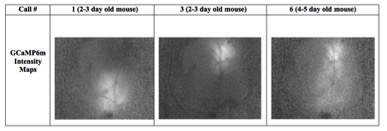
This widespread cortical GCaMP6m activation throughout the A1 PV+ neuronal network suggests that, when exposed to pup calls, female mice do not tend to differentiate among varying frequencies. Perhaps this could be due the dependency of all pups to receive this nurturing behavior, and for mothers and surrogates alike to provide it.
This suggested binary distinction of female mice¾ recognizing ‘call vs. no call,’ but not distinguishing between ‘call frequency A vs. call frequency B’¾ is one that will be further investigated in the Shea lab, as we look to hone in on the neural circuits that regulate long-term, experience-dependent plasticity in the auditory cortex.
I would like to thank my research advisor for the summer, Dr. Stephen Shea, and the directors of the Undergraduate Research Program, Dr. Anne Churchland and Kim Creteur, for providing me with this opportunity. I also thank my parents¾ Michael and Winnie Kyalwazi. Against all odds you continue to work hard and sacrifice so I have opportunities as life-changing as coming to CSHL to study neuroscience… a dream come true. Your love and support has been and will never cease to be the wind beneath my wings. It has definitely been a memorable summer for me here at Cold Spring Harbor Laboratory and I look forward to the future.
I attended the Neurofutures 2016 conference at the Allen Institute for Brain Sciences in Seattle last week. The conference that focussed on new technologies in the field and how they will drive new discoveries. I gave the opening plenary talk at the conference, a public lecture which you can see here. Following my lecture, I was part of a panel consisting of olfaction hero Linda Buck, blood flow guru (and recent marmoset pioneer) Alfonso Silva and eCog sage Jeff Ojemann. It was exciting to hear their take on the most exciting technologies in neuroscience. Some of the exciting new developments were highlighted by the panel included optogenetics, powerful transgenic animals (mice, marmosets and beyond) and high-throughput sequencing, just to name a few.

I share my colleagues’ enthusiasm for those techniques, but also held fast that these techniques must be accompanied by advances in theory to support out ability to understand the incoming data. Theoretical neuroscience has historically played a fundamental role in the field as a whole, and its importance going forward cannot be understated (I have argued for this before).
A recent paper in Neuron from Kanaka Rajan, Chris Harvey and David Tank sets out to demonstrate how relatively unstructured networks can give rise to highly structured outputs that persist on slow timescales relevant to behaviors like decision-making and working memory. Such unstructured networks seem at first like exactly the wrong thing to support stimulus-driven persistent activity. Indeed, classic work in the prefrontal cortex revealed individual neurons that respond persistently during delays, presumably support the ability of the animal to hold information in mind over that delay. In mouse posterior parietal cortex, however, it’s a different story. On a memory guided decision task published previously many individual neurons respond only very transiently, for much less time than the animal holds those memories in mind. Both that paper and the current one argue that many such neurons could fire in sequence, supporting slow-timescale memory-guided decisions even in the absence of single neurons with persistent activity.
The big steps forward in the current paper are:
- The authors demonstrated that a randomly connected network could give rise to this activity. This was an advance for a number of reasons, including the development of a new model framework called PINning. This method builds on a now classic technique, FORCE learning which generates coherent activity patterns from chaotic networks. PINing is different because only a small percentage (~12%) of synaptic weights are allowed to change. The ability of the network to capture the complex firing rates of 437 neurons when only a few synaptic weights were allowed to change is a big deal.

Network that learns by PINning; red lines are the only synapses that are allowed to change during learning to match the data.
- The paper pointed out features of the data that are incompatible with a traditional model for persistent activity, like bump attractors. This is evidence against an appealing idea (that may be present in other systems) in which a hill of activity moves around the network, driving a persistent response.
- Finally, the authors found that the network’s success relied not only on the strongly choice-selective neurons you might expect, but also on neurons that weren’t selective for the animal’s choice at all. In fact, they observed that these seemingly unimportant neurons might play a critical “conveyer belt” role that was essential in supporting more difficult decisions, especially those among many alternatives. The previous paper (and indeed many other studies) mainly excluded these neurons from analysis; an understandable choice at the time, but one that now warrants reconsideration.
There is still a challenge ahead for putative mechanisms that support slow timescale behaviors like working memory and decision-making. At the moment, there are few causal manipulations that can disrupt proposed mechanisms and demonstrate an effect on behavior. In the framework here, it would be compelling to demonstrate that changing the order of the sequence changed the behavior (admittedly no small feat!). More traditional mechanisms aren’t off the hook either: demonstrating that persistent activity at the single neuron level supports working memory likewise would be aided by precise disruption experiments. Indeed, the single neuron persistence could be epiphenomenological; the persistent working memories could be supported by some other aspect of the network. Many such manipulation experiments will be feasible in the near future.
Until then, I am excited to see a new mechanism to support slow-timescale behavior. It is counterintuitive that such network complexity can be captured by a randomly connected network, especially one in which such a small number of synapses are allowed to change.
Barbara McClintock lecture at CSHL links plants and the brain
February 11, 2016


Discoveries made by plant geneticists in the 1940s are changing our understanding of the brain. Specifically, Barbara McClintock’s (left) discovery of transposons, for which she won the nobel prize, has turned out to be important not only for understanding gene function in plants, but in brains as well. Tranposons, described by the New Yorker as “wandering snippets of DNA that hide in genomes, copying and pasting themselves at random” account for ~40% of our genome. They are likely to play a key role in normal brain function, and also might be involved in neurodegenerative diseases including ALS.
The importance of transposons for all biology inspired current CSHL graduate students and motivated them to create a lecture series named after Barbara McClintock. The first one was today, and in recognition of the role of transposons in the brain, they invited a neuroscientists, Ann Graybiel (right) from MIT, to be the first recipient. Ann’s work on the striatum has been critical for the field’s growing understanding of how incoming inputs can lead to actions, especially ones that are reinforced and become habitual. Her emerging work is especially exciting as her lab is leveraging modern techinques to specifically measure and manipulate classes of cells within the striatum to understand their role in different behaviors and decisions.
To commemorate the creation of this new lecture series and its first recipient, neuroscientists from around New York gathered to honor Ann and attend her talk. Researchers focussing on decision-making, attention, vision and auditory processing came together and some lively discussions ensued! It was a lot of fun to show the setups in the my lab to this crew, which included Jackie Gottlieb, Yael Niv, Heather Read and Ariana Maffei, and we realized many links between our collective research programs that I hope will lead to new collaborations down the line.

I am happy to announce another post by a guest blogger. This time, its Sashank Pisupati, a new graduate student in my lab.
Last week, our lab read a paper by Ramon Reig & Gilad Silberberg titled “Multisensory Integration in the Mouse Striatum”. While studies of multisensory integration have focussed largely on cortical structures and the superior colliculus, this study adds to a growing body of evidence that the striatum may play a key role in this process. Striatal medium-spiny neurons (MSNs) are known to receive convergent projections from multiple sensory cortices, but relatively few studies have reported multisensory responses in these cells.
Here, the authors set out to test whether individual MSNs integrated visual (LED flashes) and tactile (whisker stimulation with air puffs) stimuli in anesthetized mice. In order to observe such synaptic integration, they performed whole-cell patch clamp recordings from striatal neurons. They targeted regions of striatum receiving projections from primary visual (V1) and somatosensory (S1) cortex, as identified by anterograde tracing using BDA.
They found sub-threshold responses to whisker stimulation (purple trace) in all the neurons they recorded from, which were modulated by stimulation strength. More interestingly, in the dorsomedial striatum a subset of these neurons were also responsive to visual stimuli (green trace), with slightly longer peak response latencies. They then presented visual and tactile stimuli together at various relative delays, and observed multisensory responses in these cells (orange & black traces) that were sublinearly additive i.e. less than the linear summation (grey traces) of the visual and tactile responses. Moreover, the peak multisensory response was maximal when the onsets/peaks of the unisensory responses were aligned, suggesting that the neurons summated congruent synaptic inputs.

These findings of multisensory cells in the mouse striatum corroborate similar reports from extracellular recordings in the striatum of rats and cats, and complement them by offering a valuable glimpse of sub-threshold activity. The sub-linear additivity described here contrasts with the super-linear additivity of firing rate responses often emphasized in studies of the superior colliculus.
One of the questions that remained at the end of our discussion, was how this result fit into models of multisensory integration such as divisive normalization4, or Bayes-optimal cue combination. While classical approaches have emphasized the degree of additivity of the unisensory responses, these models make strong predictions about how the weights assigned to each unisensory response in the summation change in accordance to the reliability of that sensory modality4. For example, we expect the contribution of a visual flash to the summation to decrease for weaker, less reliable flashes.
One could test this prediction in the author’s current setup by simply varying the stimulus strength for each modality during multisensory presentation. Combined with the power of the patch clamp approach, this could yield further insight into the sub-threshold computations being performed by these neurons, and we hope to see more such work in the future!
Neural perturbations can have minimal effects even in structures that are causal for behavior
November 9, 2015
 This post is written by guest blogger, Matt Kaufman, a postdoc in my lab (left).
Last week our lab read a recent Neuron paper out of the Brody lab, by Kopec, Erlich, Brunton, Deisseroth & Brody, titled “Cortical and subcortical contributions to short-term memory for orienting movements.” This paper continues with that lab’s recent strategy of using optogenetics to briefly inactivate brain areas during decision making.
The experiments were straightforward. They trained rats to judge whether a click train was faster or slower than 50 Hz, then used optogenetics (eNpHR3.0) to inactivate either the Frontal Orienting Fields (FOF) or superior colliculus (SC) on one side of the brain at different points in the trial. This allowed Kopec et al. to see when these areas contributed to making the decision. The key experimental finding was that the rats’ decisions were most biased when either FOF or SC was silenced during the stimulus, a little less biased when silenced early in the subsequent delay, and less biased still when silenced late in the delay. Decisions were essentially unaffected when silencing was performed during the response period.
This finding is initially surprising, because tuning in the FOF increases over the course of the trial (as known from previous studies). They argue, however, that this seeming mismatch makes sense in the context of an attractor dynamics model (below). Since the evidence from the stimulus is not fluctuating in this task, the animal should be able to make his decision quickly. The increasing tuning might be due to attractor dynamics that amplify the tuning with time, while perturbations should mostly impact decisions before the neural activity has had time to settle in an attractor. Additional comparisons, including inactivating both areas together and comparing hard vs. easy trials, quantitatively fit their simple attractor model.

This study forms an interesting contrast with their paper from earlier this year, Hanks et al. 2015 in Nature. There, they took a similar approach but with a temporal integration task. In that task, the FOF was only critical at the end of such a stimulus. This again makes sense; you don’t want attractor dynamics if you need to integrate instead.
The question on many of our minds was: do these areas “really” exhibit attractor dynamics? On further reflection, though, this is a bit like asking whether the planets follow Newton’s laws. What I mean by that is: neurons, like orbiting planets, aren’t solving equations. Dynamical models, like Newton’s equations, are a mathematical description of how the system behaves over time. But if a model is an easy way to think about a system, and makes intuitive, useful predictions that hold up experimentally, then the model does useful work.
Many questions remain unanswered, of course. In terms of separation of function, are FOF and SC really doing the exact same thing? Are there other tasks where they would function very differently? Regarding dynamics, how does the system learn to produce these attractor dynamics? Since the FOF can apparently be trained to produce different dynamics in animals trained on different tasks, can it support either computation in an animal trained on both tasks? If so, how would it switch its dynamics? We’ll look forward to the next installment.
